Medical Image Formats: DICOM, NIfTI & Analyze
Objective
The data layer under every medical imaging system
Hospitals do not store PNGs. Scanners write DICOM with patient and acquisition metadata, neuroimaging research uses NIfTI volumes with spatial orientation, and legacy archives hold Analyze header/image pairs.
This experiment loads each format programmatically, inspects its array shape and dimensionality, and renders a representative slice — the first step of any real-world medical imaging or AI pipeline.
Tools & Data
Formats Handled
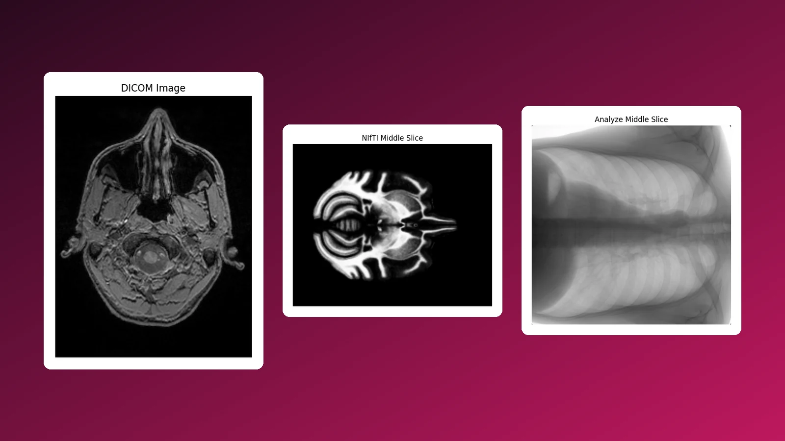
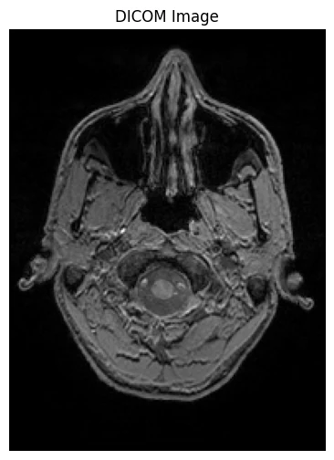
The hospital standard: pixel data plus patient and imaging metadata. Read a brain MR slice and rendered its pixel array.
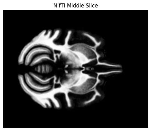
3D/4D neuroimaging volumes with orientation. Loaded a (168 × 206 × 128 × 1) brain volume and displayed the middle axial slice.
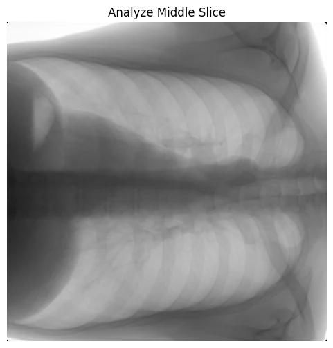
Legacy split header/image format. Loaded a (440 × 440 × 1) chest X-ray and displayed it.
Figures
DICOM brain MR · NIfTI middle slice · Analyze chest X-ray
Why It Matters
Clinical data engineering
Any medical AI product, PACS integration or research pipeline lives or dies on correct handling of these formats — orientation, dimensionality and metadata. Being comfortable at this layer connects my software engineering work, such as healthcare platforms like Medisheba, with clinical imaging data.
Coursework for the Medical Imaging Laboratory (BME-3202), B.Sc. Biomedical Engineering, Islamic University.