MRI Reconstruction from Multi-Coil k-Space
Objective
From scanner frequencies to a readable image
An MRI scanner never records a picture. It samples k-space: the spatial-frequency content of the anatomy, collected line by line through magnetic field gradients and recorded separately by each receiver coil.
The goal of this experiment was to take raw multi-coil k-space straight from a scanner dataset and reconstruct the final diagnostic image myself, understanding every step a clinical reconstruction pipeline performs.
Tools & Data
Reconstruction Pipeline
Each stage implemented and visualised
-
Load raw k-space
Read the HDF5 volume with shape (slices, coils, height, width) and select the middle slice for reconstruction.
-
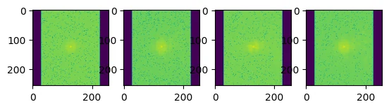
Inspect k-space per coil
Display log-magnitude k-space for four coils: the bright centre holds low frequencies (contrast and brightness), the periphery holds edges and fine detail.
-
Inverse Fourier transform
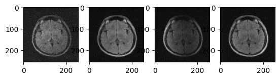
Convert each coil's k-space to a PyTorch tensor and apply a centred inverse 2D FFT, producing a complex-valued image per coil.
-
Magnitude and phase
Separate each complex pixel into magnitude (the visible brightness) and phase, keeping magnitude for display.
-
Coil combination
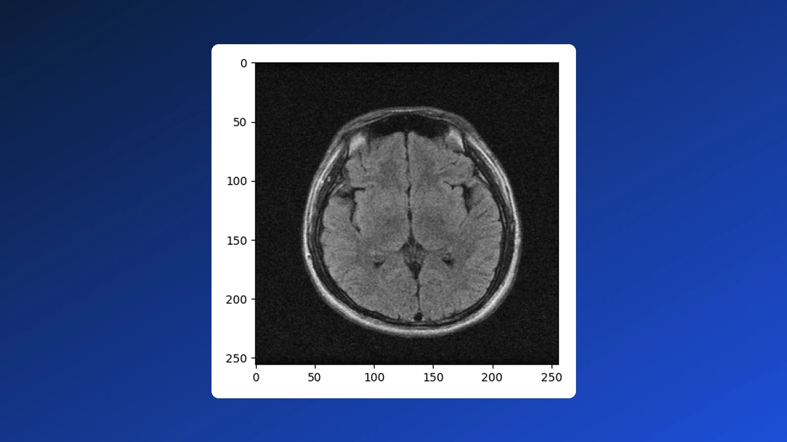
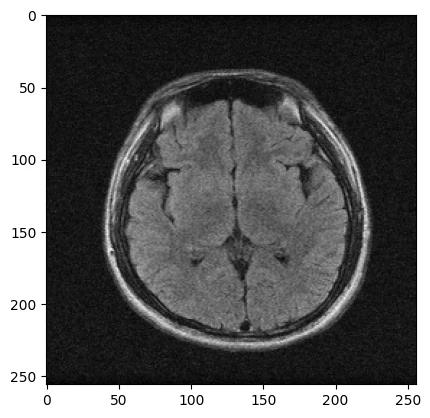
Fuse the coil images with Root-Sum-of-Squares so every coil's regional sensitivity contributes to one uniform image.
Coil-by-Coil Reconstruction
Key Concepts Demonstrated
Centre of k-space governs contrast; outer regions carry edges — completeness of sampling decides image quality.
The inverse FFT yields real and imaginary parts per pixel; magnitude and phase are derived from them.
Each receiver coil sees a different region strongly; combining them recovers the full field of view.
Per-pixel square root of summed squared coil magnitudes — the standard coil-combination baseline.
Figures
k-space per coil · coil images · final RSS reconstruction
Why It Matters
The foundation of accelerated and AI-driven MRI
Every modern accelerated-MRI technique — parallel imaging, compressed sensing and deep-learning reconstruction such as the fastMRI challenge models — starts from exactly this pipeline. Being fluent in raw k-space, complex-valued data and coil combination is the prerequisite for building or validating learned reconstruction models.
Coursework for the Medical Imaging Laboratory (BME-3202), B.Sc. Biomedical Engineering, Islamic University. Data: M4Raw dataset. Library: fastMRI (Meta AI / NYU Langone).